Friday, October 31, 2008
2008.10.31
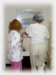
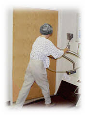
Linda learned at 8:30 am she could leave and she checked out of Carolinas Medical Center at 12:15. Eye issue remains.
Appointments to remove stitches and staples for Wednesday 11/05 at 10am at Dr Asher's office, with PT on Thursday 11/06 at 9:00 am be there at 8:30 at CMC main, followup with Dr Asher on Monday 11/24 at 10:45 AM.
I was in charge of dinner tonight - Wolfman Pizza, a BLT and Baked Potato Pizza, super if you have not tried them. Tomorrow we do grocery shopping and get some healthy foods. Marlene has cleaned out Linda's refrigerator to start over.
Someone is to be with Linda 24 hours a day for a while and I am on duty tonight. Tomorrow Marlene is on duty as Marlene and Linda decided I could have the weekend off to watch football nonstop - thanks!
Thursday, October 30, 2008
2008.10.30
Linda might go home on Friday, She is doing well but eye issues continue.
We saw Dr Asher and folks from Physical Therapy today. Looks like she will not need the PT in hospital and might do on out patient basis, if needed. She obviously is progressing well and fast, except for the eyes. We were told to give it a couple of weeks.
2008.10.29
Dr Asher said this morning the double vision was normal and from swelling and they are doing some Physical therapy with it.
We are working into a schedule where Marlene goes in the morning and I go after lunch until 9 or 10. That works better for me as I am not a good getting up early in the morning person, rather a stay up past midnight guy.
Tuesday, October 28, 2008
2008.10.28
Another good day. Linda had most of her tubes removed today, she was sitting up and some walking, and her swelling way down. Still double vision. She is enjoying flowers, calls, and visits and even a book on tape someone brought by. Thanks for your kindness.
Dr. Howell was by and as he was leaving said he needed to go and visit some sick people! Thanks for coming by and for your prayer of thanksgiving and encouragement.
The Physical Therapist was by and did some preliminary checking.
First time I saw the staples and stitches on the top of her head. Amazing. Her hair will cover the area after they are removed.
Monday, October 27, 2008
2008.10.27
Linda's room number as you know is 9910 (Carolinas Medical Center, 1000 Blythe Blvd, charlotte nc 28203) and now we know her telephone > 704 355 6801. Try again if busy.
She had a very good day. I had just left seeing her this morning and she called me excited that sister Marlene had washed on her eyes and, lo, they opened and she can see. She had some double vision but both eyes work. By the end of the day she said one eye was clear with the other not so clear and she was not seeing as many doubles.
One doctor Tuesday said they "got all of it" in the surgery. Previously they were qualifying it by saying there were 4,000 somethings we were dealing with.
First thing this morning she was asking what the stock futures were so she is staying in touch. (I said I did not know as I dreaded knowing, the way things are going in the markets.)
Rev. Shane Page (MPUMC Minister of Evangelism and Involvement) came by and Linda appreciated that. He looked neat in his collar and blue jeans. He shared that Carol Douglas was in the same hospital in room 10-920 after visiting the Emergency Room Sunday night.
By end of day the swelling was down but still there. Also color was better.
Sunday, October 26, 2008
2008.10.26
The really good news is that a draining tube which had not been working and causing some concern was "fixed" by her Dr. Asher. Previously he had said he was unable to do it but by some method today he did. He said it made him a happy man. Otherwise the draining was to have to be into the face area and then naturally dissipate.
Linda was moved during the night from Intensive Care into room 9910. Visiting hours are open and she is doing ok with visitors. She skipped Progressive Care, so perhaps that is a good sign.
Her eye area continues to swell and she still has not opened her eyes. Putting cold water there to help .Eating regular food and no more IV. I read the headlines to her from the paper and shared what was going on in the world. We "watched" the end of the panther game with my updating her during the comeback. She still is upset about the terrible game at Tampa.
Rev. Bill Roth (MPUMC Minister of Congregational Care) came by today when I was not there and Linda was asleep. He said a prayer and left. When I came I saw his card and later Linda said that Bill Roth had said a beautiful prayer so maybe she was playing possum. (Mystery solved, Linda said Bill Roth also called and that is when she heard his prayers.)Thanks Bill. Which reminds me, she appreciates your prayers and support
On a sad note, Bertha's husband had a stroke yesterday (while with about 15 of their family in Florida for a family reunion). He was flown back to Charlotte this morning at about 4:30 am. They are awaiting MRI results but right now he cannot or is not being allowed to eat through the mouth. Loss of movement on one of his sides. Speech not working but he knows what is going on around him , waving goodbye for example when I left. Bertha (Gerald) is a lady who does house cleaning at my home and at Linda's several times a month. We were counting on her so that might be modified going forward as she deals with him. He is at Presbyterian Healtcare ,200 Hawthorn Lane, Charlotte NC 28233 (hospital # is 704 384 4000) room 479 and his name is John Gerald Jr. Their son is a minister of Gethsamine Baptist Church.
Saturday, October 25, 2008
2008.10.25
Good day. Her nurse said Linda was doing better than 95% of patients in this situation. All day she was able to communicate and knew it was Saturday, remembered I told her UNC won, said she was sorry I did not get to go to game, and generally was in touch. She talked of wanting to go home. She does have medication. She has been up a couple of times but that is hard. She asked if I were going to Sunday School tomorrow (10/25). I understand during tonight of 10-25/26 she probably will move from Intensive Care (room 9616) to Progressive Care or regular care. Yesterday she could open her eyes better then today. I asked if she could look at me and she said she couldn't open her eyes. The swelling is and was to be expected. We should know her room number in a day or so. She knows about cards, calls, and visits , etc of people and of your support. Feel free to contact me in any way. Thanks.
Friday, October 24, 2008
2008.10.24
Surgery lasted about 11 1/2 hours beginning at 8:12 am. Dr Asher talked with us for over 40 minutes after surgery.
In my attempting to make a long story short , I asked Dr. Asher what to say to friends without medical degrees.
He said the tumor was benign but extremely complex and involved both frontal lobes. The procedure took longer than we anticipated, but he was pleased with how things turned out.
There are many more details of the risks remaining and the time involved going forward. Linda came through a tough day. She has been brave the whole time. She was able to tell her birthday and other info a couple of hours after surgery. She will be in Intensive Care a day or so . .and will be sleeping a lot. Swelling is expected to increase for a day or so.
Thursday, October 23, 2008
Wednesday, October 22, 2008
2008.10.22
Our dear friend, Linda Myers, has surgery Friday October 24 at 7:30 am at Carolinas Medical Center in Charlotte. She has a meningioma brain tumor located near the skull on the front high forward. The surgeon is Dr Tony (Anthony L.) Asher from the Carolina Neurosurgery and Spine Associates. Prognosis is good but there are risks. Surgery is expected to last 4 to 5 or 6 or 8 hours. She will be in intensive care for 2-3 days, hospital 7-10 days, rehab 2-3 days, recovery over weeks and maybe months. It is near pituitary gland, optic nerve, etc. Such tumors mainly are benign. This type is slow growing for years, giving time to catch and treat. It is the size of a flat tennis ball. Dr Asher says non-surgery is not an option as it eventually is life threatening. She is taking steroids to reduce the inflammation around the tumor. The steroids are giving her more trouble with her stomach, etc, than the tumor. There is no pain now. She is able to do about all she usually does on a daily basis and would have been at Sunday School last week except for family from out of town celebrating her birthday a few weeks early. Several factors, including loss of energy and sense of smell, led to the MRI October 02, 2008, which identified the tumor. She is in good spirits and wants to get this over. Her mailing address is 4739 Hedgemore Drive Unit T, Charlotte NC 28209 . She is not doing emails right now. She does not know her room yet or CMC address (other than 1000 Blythe Blvd, 28203). She asks for your prayers and moral support. (2008.10.22)
Tuesday, October 21, 2008
Meningioma
For friends of Linda who want to know
Meningioma
From Wikipedia, the free encyclopedia
| Meningioma Classification and external resources | |
| A contrast enhanced CT scan of the brain, demonstrating the appearance of a Meningioma. | |
| ICD-10 | C70, D32 |
| ICD-9 | 225.2 |
| ICD-O: | 9530 |
| OMIM | 607174 |
| DiseasesDB | 8008 |
| eMedicine | neuro/209 radio/439 |
| MeSH | D008579 |
Meningiomas are the most common primary tumor of the central nervous system, arising from the arachnoid "cap" cells of the arachnoid villi in the meninges.[1] These tumours are usually benign in nature; however, they can be malignant.[2]
Contents |
Causes
Most cases are sporadic while some are familial. Persons who have undergone radiation to the scalp are more at risk for developing meningiomas.[3]
The most frequent genetic mutations involved in meningiomas are inactivation mutations in the neurofibromatosis 2 gene (merlin) on chromosome 22q. Other possible genes/loci include: MN1[4] PTEN[5] an unknown gene at 1p13[6]
MRI - Magnetic resonance imaging
For Linda's friends who want to know:
Magnetic resonance imaging
From Wikipedia, the free encyclopedia

Magnetic resonance imaging (MRI), or nuclear magnetic resonance imaging (NMRI), is primarily a medical imaging technique most commonly used in radiology to visualize the structure and function of the body. It provides detailed images of the body in any plane. MRI provides much greater contrast between the different soft tissues of the body than computed tomography (CT) does, making it especially useful in neurological (brain), musculoskeletal, cardiovascular, and oncological (cancer) imaging. Unlike CT, it uses no ionizing radiation, but uses a powerful magnetic field to align the nuclear magnetization of (usually) hydrogen atoms in water in the body. Radiofrequency fields are used to systematically alter the alignment of this magnetization, causing the hydrogen nuclei to produce a rotating magnetic field detectable by the scanner. This signal can be manipulated by additional magnetic fields to build up enough information to construct an image of the body.
MRI is a relatively new technology, which has been in use for little more than 30 years (compared with over 110 years for X-ray radiography). The first MR Image was published in 1973[1] and the first study performed on a human took place on July 3, 1977.[2]
Magnetic resonance imaging was developed from knowledge gained in the study of nuclear magnetic resonance. In its early years the technique was referred to as nuclear magnetic resonance imaging (NMRI). However, as the word nuclear was associated in the public mind with ionizing radiation exposure it is generally now referred to simply as MRI. Scientists still use the term NMRI when discussing non-medical devices operating on the same principles. The term Magnetic Resonance Tomography (MRT) is also sometimes used. One of the contributors to modern MRI, Paul Lauterbur, originally named the technique zeugmatography, a Greek term meaning "that which is used for joining".[1] The term referred to the interaction between the static, radiofrequency, and gradient magnetic fields necessary to create an image, but this term was not adopted.
Contents
[hide]CAT Scan/CT Scan - what is it
For Linda's friends who want to know what a CatScan is:
Computerized Axial Tomography
(CAT Scan/CT Scan)
Medical Author: Melissa Conrad Stoppler, MD
Medical Editor: William C. Shiel, Jr, MD, FACP, FACR
- What is a CT scan?
- Why are CT scans performed?
- Are there risks in obtaining a CT scan?
- How does a patient prepare for CT scanning, and how is it performed?
- CT Scan At A Glance
- Patient Discussions: Ct Scan - Helped With Your Diagnosis
What is a CT scan?
A computerized axial tomography scan is an x-ray procedure that combines many x-ray images with the aid of a computer to generate cross-sectional views and, if needed, three-dimensional images of the internal organs and structures of the body. Computerized axial tomography is more commonly known by its abbreviated names, CT scan or CAT scan. A CT scan is used to define normal and abnormal structures in the body and/or assist in procedures by helping to accurately guide the placement of instruments or treatments.
A large donut-shaped x-ray machine takes x-ray images at many different angles around the body. These images are processed by a computer to produce cross-sectional pictures of the body. In each of these pictures the body is seen as an x-ray "slice" of the body, which is recorded on a film. This recorded image is called a tomogram. "Computerized Axial Tomography" refers to the recorded tomogram "sections" at different levels of the body.
Imagine the body as a loaf of bread and you are looking at one end of the loaf. As you remove each slice of bread, you can see the entire surface of that slice from the crust to the center. The body is seen on CT scan slices in a similar fashion from the skin to the central part of the body being examined. When these levels are further "added" together, a three-dimensional picture of an organ or abnormal body structure can be obtained.
Echo Cardiogram
| Echo Stress Test |
How do I prepare for the test?
How is the test performed
How long does it take?
How safe is it?
What is the reliability of the test?
How quickly will I get the results?
Show me a panoramic view of the Echo Stress lab
How does Stress Echo work? Patients with coronary artery blockages may have minimal or no symptoms during rest. However, symptoms and signs of heart disease may be unmasked by exposing the heart to the stress of exercise. During exercise, healthy coronary arteries dilate (develop a more open channel) than an artery with a blockage. This unequal dilation causes more blood to be delivered to heart muscle supplied by the normal artery. In contrast, narrowed arteries end up supplying reduced flow to it's area of distribution. This reduced flow causes the involved muscle to "starve" during exercise. The "starvation" may produce symptoms (like chest discomfort or inappropriate shortness of breath), EKG abnormalities and reduced movement of the heart muscle. The latter can be recognized by examining the movement of the walls of the left ventricle (the major pumping chamber of the heart) by Echocardiography.
In the animation shown above, the left hand panel, marked "Resting" shows normal movement of the septum (the muscle partition between the right and left ventricles (RV and LV, respectively) while the patient is resting. The animated echo on the right ("Exercise") shows that movement of the septum is markedly reduced immediately following stress. Such findings would indicate a blockage in the artery supplying the partition of the heart and the front portion of the left ventricle (both these areas are supplied by the LAD or left anterior descending coronary artery).
How is a Stress Echo performed? An Echo Stress can be obtained in a physician's office or in the hospital. Imaging tests are generally obtained when a physician wishes to confirm or rule out the presence of coronary artery disease. A Stress Echo is also performed in patients who have disease involving the heart muscle or valve, or if a patient is having inappropriate shortness of breath and a cardiac cause is suspected.
The patient is brought to the Echo laboratory where a "resting" study is performed. This provides a baseline examination and demonstrates the size and function of various chambers of the heart. Particular attention is paid to the movement of all walls of the left ventricle (LV). Similar to a regular echo test, sticky patches or electrodes are attached to the chest and shoulders and connected to electrodes or wires to record the electrocardiogram (EKG or ECG). The EKG helps in the timing of various cardiac events (filling and emptying of chambers).
A colorless gel is then applied to the chest and the echo transducer (as described in the Echocardiogram section) is placed on top of it. The echo technologist then makes recordings from different parts of the chest to obtain several views of the heart. You may be asked to move form your back and to the left side. Instructions may also be given for you to breathe slowly or to hold your breath. This helps to obtain higher quality pictures. The images are constantly viewed on the monitor. It is also recorded on photographic paper, on videotape and on a computer disk.
  |
12 leads of the EKG are recorded on paper and the blood pressure is taken. Exercise is then initiated using a treadmill (most common) or a stationary bicycle. In patients who are unable to complete a high level of exercise because of physical limitations, stress to the heart is provided by pharmaceutical or chemical stimulation of the heart. Stress Echo is made up of three parts: A resting Echo study, Stress test, and a repeat Echo while the heart is still beating fast.
Exercise stress testing usually employs the "Bruce" or a similar protocol, as described in the Regular Stress Test section. Exercise is started at a slower "warm-up" speed. The speed of the treadmill and it's slope or inclination is increased every 3 minutes. The treadmill is abruptly stopped when the patient exceeds 85% of the target rate (based upon the patient's age). Exercise may be stopped earlier if the patient develops alarming symptoms (chest discomfort, marked shortness of breath, weakness, dizziness, etc.), if there is dangerous elevation or drop in the blood pressure, significant EKG changes or a potentially dangerous irregular heart rhythm. Please remember that you have a physician in attendance (although an experienced assistant may perform the test if the physician is tied up with an emergency). The above problems are uncommon and you are far safer if they occur in the presence of an experienced medical team rather than having them happen while you are exercising in a spa, jogging, or running up a flight of office stairs.
EKG recordings are made during every minute of exercise and then again after exercise is stopped. The blood pressure is recorded at three minute intervals during exercise and then again at rest.
Immediately after stopping the treadmill, the patient moves directly to the examination table and lays on the left side. The Echo examination is immediately repeated. Images are stored and then played back by the computer. A video clip of multiple views of the resting and exercise study are compared side-by-side. They are analyzed by the physician. Normally, one expects an increased EF or ejection fraction (a measure of how well the heart is pumping). Also, the LV walls do not show any exercise-induced abnormal movement. In contrast, a drop in EF and/or a new wall motion abnormality is an indicator of disease.
Preparing for the Echo Stress Test: The following recommendations are "generic" for all types of cardiac stress tests:
- Do not eat or drink for three hours prior to the procedure. This reduces the likelihood of nausea that may accompany strenuous exercise after a heavy meal. Diabetics, particularly those who use insulin, will need special instructions from the physician's office.
- Specific heart medicines may need to be stopped one or two days prior to the test. Such instructions are generally provided when the test is scheduled.
- Wear comfortable clothing and shoes that are suitable for exercise.
- An explanation of the test is provided and the patient is asked to sign a consent form.
How long does the entire test take? A patient should allow 1 1/2 to 2 hours for the entire test, including the preparation, echo imaging and stress test.
How safe is a Stress Echo test? There are no known adverse effects from the ultrasound used during Echo imaging. The risk of the stress portion of the test is rare and similar to what you would expect from any strenuous form of exercise (jogging in your neighborhood, running up a flight of stairs, etc.). As noted earlier, experienced medical staff is in attendance to manage the rare complications like sustained abnormal heart rhythm, unrelieved chest pain or even a heart attack. These problems could potentially have occurred if the same patient performed an equivalent level of exercise at home or on a jogging track.
What is the reliability of Stress Echo? If a patient is able to achieve the target heart rate and if the ECHO images are of good technical quality, a Stress Echo is capable of diagnosing important disease in more than 85% of patients with coronary artery disease. Also, it can exclude important disease in more than 90% of cases when the test is absolutely normal.
How quickly will I get the results and what will it mean? The physician conducting the test will be able to give you the preliminary results before you leave the Stress Echo laboratory. However, the official result may take a few days to complete. The results of the test may help confirm or rule out a diagnosis of heart disease. In patients with known coronary artery disease (prior heart attack, known coronary blockages, previous treatment with angioplasty, stents or bypass surgery, etc.), the study will help confirm that the patient is in a stable state, or that a new blockage is developing. The results may influence your physician's decision to change your treatment or recommend additional testing such as cardiac catheterization.
The panoramic view (below) shows a patient undergoing the treadmill and Echocardiography portions of the stress test (combined into a single picture).
You may also pan left and right by clicking and draggging your mouse within the panoramic picture.

